
Figure 1.
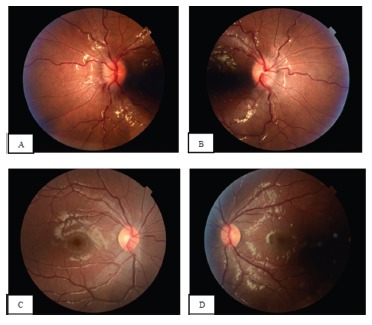
Fundus photography of the right (A) and left (B) eyes showing bilateral papilledema with optic nerve head elevation, peripapillary hemorrhages and vessel tortuosity.
Fundus photography of the right eye (C) and left eye (D) 4 months after treatment showing significant improvement of disc edema. Some peripapillary nerve fiber layer opacification remains.