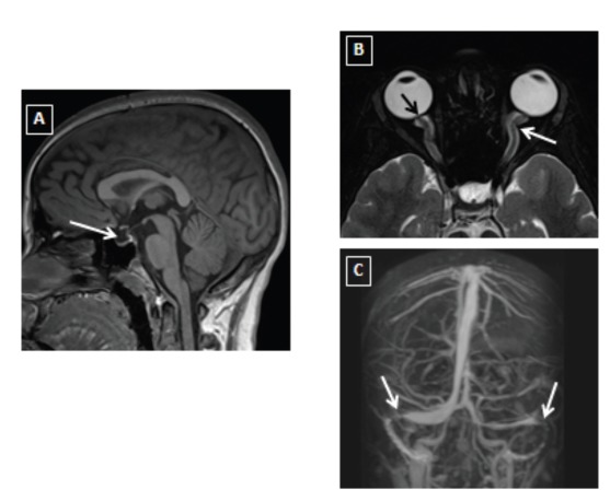
Figure 2.

Brain MRI (A, sagittal T1WI) showing partial empty sella (white arrow). Axial T2 fat saturation for orbits (B) showing flattening of posterior globe and prominence of the optic nerve head (black arrow) as well as tortuosity of optic nerve and prominent perioptic nerve sheath (white arrow). MRV (C) showing focal narrowing of bilateral distal transverse sinuses (white arrow).
