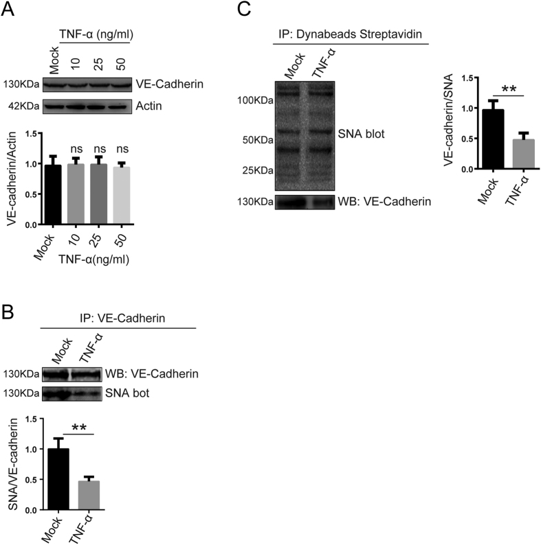
Figure 3. VE-Cadherin α-2, 6 sialylation levels were decreased under proinflammatory conditions.
(A) VE-cadherin levels in EA.hy926 cells treated with TNF-α (0, 10, 20, and 50 ng/ml) for 24 h were examined via western blot assays. (B) VE-Cadherin α-2, 6 sialylation levels in EA.hy926 cells were decreased after TNF-α (50 ng/ml) treatment for 24 h. VE-cadherin was immunoprecipitated with anti-human VE-cadherin polyclonal antibodies. VE-cadherin immunoprecipitates were analyzed via SNA blotting using biotinylated SNA and HRP-labeled Streptavidin. (C) Proteins modified by α-2, 6 sialylation in EA.hy926 cells were recognized via biotinylated SNA blotting and then precipitated with Dynabeads Streptavidin. VE-Cadherin levels in the precipitates were analyzed by western blotting using antibodies against VE-cadherin. The data are the mean ± SD of three independent assays (**p < 0.01).

