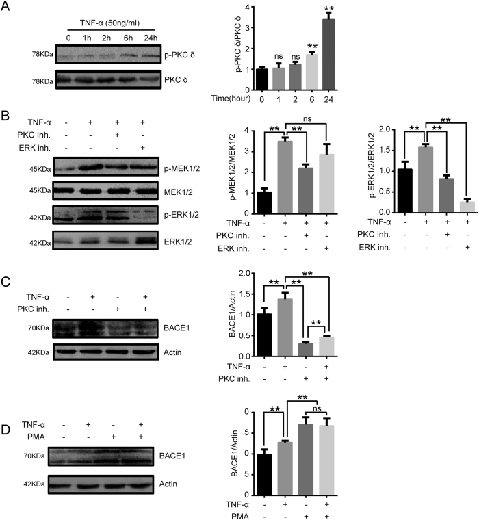
Figure 6. BACE1 expression induced by the proinflammatory cytokine TNF-α is PKC-dependent.
(A) EA.hy926 cells were treated with TNF-α (50 ng/ml) for the indicated time periods. Western blotting was conducted to characterize phospho-PKC δ levels. Quantification of normalized densities for p-PKC δ and PKC δ is shown. The graphs represent the relative activity of these kinases for three independent experiments. ns, ns, not significant versus untreated cells. **p < 0.01 versus untreated cells. (B) EA.hy926 cells were stimulated with TNF-α (50 ng/ml) with or without PKC inhibitor (10 μM) or ERK inhibitor (20 μM) for 24 h, and MEK1/2, phospho-MEK1/2, ERK1/2 and phospho-ERK1/2 levels were analyzed via western blotting assays. ns, not significant. **p < 0.01. (C,D) Effect of the PKC pathway on BACE1 expression in EA.hy926 cells. The cells were pretreated with a PKC activator (10 μM) for 6 h or PKC inhibitor (10 μM) for 30 min and then incubated with TNF-α (50 ng/ml) for 24 h. BACE1 expression levels in EA.hy926 cells were analyzed via western blotting. ns, not significant. **p < 0.01.

