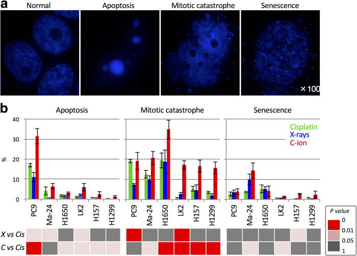
Figure 4. Clonogenic cell death induced by cisplatin, X-rays, or carbon ions.
Cells were treated with X-rays (4 Gy), carbon ions (4 Gy), or cisplatin (1 μM for 24 h) and then stained with DAPI 72 h later. Apoptosis, mitotic catastrophe, and senescence were determined according to characteristic nuclear morphologies (see “Materials and Methods” for definitions). (a) Representative images showing the nuclear morphology of normal cells and cells undergoing apoptosis, mitotic catastrophe, and senescence. Images of untreated Ma-24 cells (normal) and cells treated with cisplatin (undergoing apoptosis, mitotic catastrophe, or senescence) were taken using a DeltaVision (GE Healthcare, Little Chalfont, UK) and deconvoluted and processed using SoftWoRx (GE Healthcare)35. (b) Mode of clonogenic cell death in cells treated with X-rays or carbon ions plus cisplatin. In the lower panels, the statistical significance of differences in the levels of apoptosis, mitotic catastrophe, or senescence induced by X-rays (X) or carbon ions (C) plus cisplatin (Cis) is indicated by different colors.

