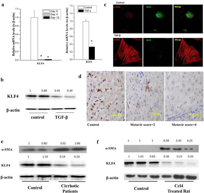
Figure 4. KLF4 was down-regulated in activated HSCs and cirrhotic liver tissues.
(a) The mRNA level of KLF4 dramaticlly decreased in the process of spontaneous HSC activation (left pannel) and primary HSCs treated with 10 ng/mL of TGF-βfor 24 hours (right pannel). (b) KLF4 protein level was significantly decreased in HSCs by treating primary HSC with 10 ng/mL TGF-β for 24 hours. (c) TGF-β down-regulated KLF4 while upregulatedα-SMA in rat primary HSCs. KLF4 and α-SMA were detected by immunocytochemical staining and pictures were taken with a confocal microscopy. Rat primary HSCs were treated with 10 ng/mL TGF-β for 4 hours. (d) KLF4 was significantly suppressed in cirrhotic liver of patients as compared with the healthy controls (scored by immunohistochemistry studies, n = 16). (e) KLF4 was significantly down-regulated, whileα-SMA was obviously up-regulated in human cirrhotic liver tissues compared with the healthy controls. (f) KLF4 was elevated in CCl4 induced cirrhotic liver of rats. All experiments were repeated thrice with triplicate samples in each experiment. The relative value of target mRNA/protein to β-actin was set as 1 in the control. Data are presented by mean ± standard deviation. *p < 0.05; **p < 0.01.

