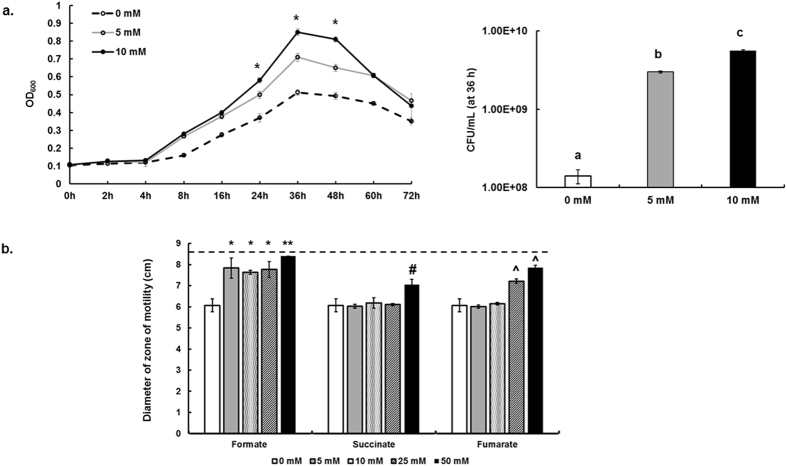
Figure 2.
(a) Growth of C. jejuni 81–176 in Mueller-Hinton broth supplemented with different concentrations of formate. Growth was determined by measuring OD600. CFU numbers were quantified at 36 h. Asterisks and different letters indicate statistically significant differences between all treatments (P < 0.05). (b) Motility of C. jejuni 81–176 in Mueller-Hinton semi-solid (0.4%) agar supplemented with different concentrations of formate, succinate, and fumarate, respectively. The diameter of zone of motility was measured after 48 h under microaerobic conditions. Note that the diameter of the Petri dish was approximately 8.5 cm, which precluded assessment of the diameter beyond 8.5 cm. Horizontal dashed line indicates the maximum limit of quantification in this assay. Symbols indicate statistically significant differences (P < 0.05) in comparison to the control (0 mM). **Denotes the most significant increase (P < 0.05) in motility across all treatments.

