Figure 3.
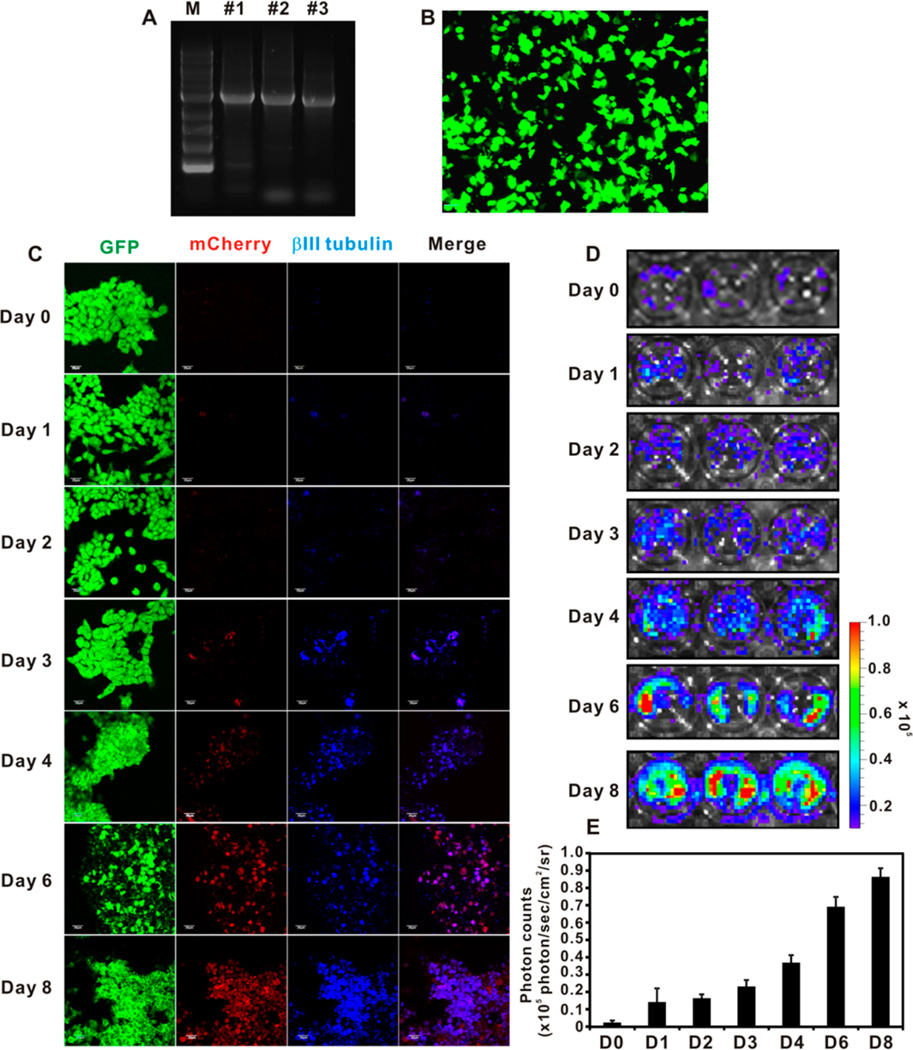
Construction of bicistronic vector with tubb3 promoter driven reporter gene. (A) Gel electrophoresis of the cloned tubb3 promoter from three independent brain tissue extracts. The #2 clone was used in the entire study. (B) Fluorescence image of lentivirus transduced NE-4C NSCs with tubb3 promoter driven fLuc reporter gene and independent EF1α driven GFP reporter gene. Scale bar 50 µm. (C) Confocal laser scanning microscopy imaging of NE-4C NSC differentiation by 100 nM RA-loaded nanovehicle over 8 days. βIII tubulin was stained by Cy5.5-conjugated secondary antibody. The GFP and mCherry were endogenously expressed fluorescence proteins in transduced NSCs. Scale bar = 50 µm. (D) Bioluminescence imaging (BLI) of tubb3 promoter driven fLuc expression in transduced differentiating NSCs stimulated by 100 nM RA-loaded nanovehicle over 8 days. (E) Analysis of BLI results (n = 3).

