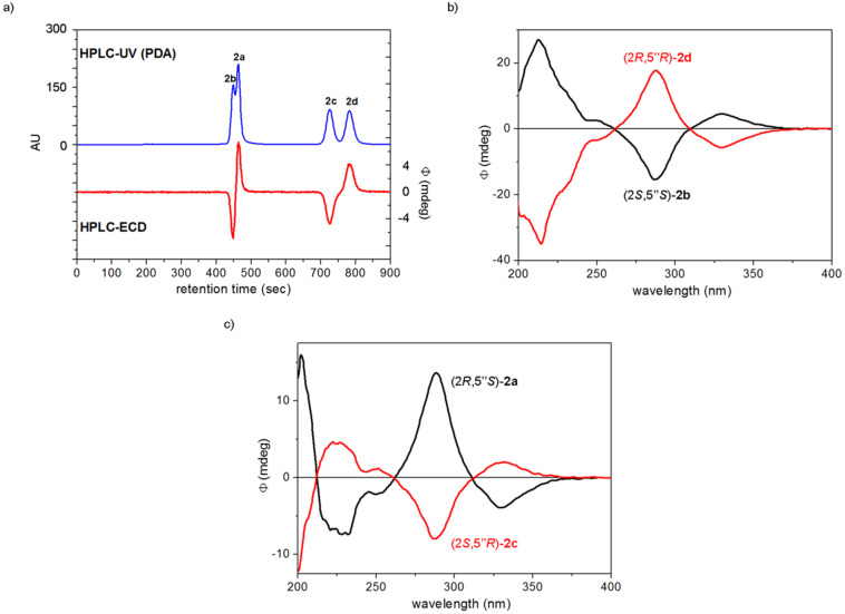
Figure 2.
a) Chiral HPLC–UV and HPLC–ECD traces of dracocephins A (2a–d) using Chiralpak IC column with the eluent MeCN/2-propanol/TFA 97:3:0.1 monitored at 290 nm. b) HPLC–ECD spectra of the first [black: (2S,5”S)-2b or dracocephin A2] and fourth eluted [red: (2R,5”R)-2d or dracocephin A4] stereoisomers of dracocephins A. c) HPLC–ECD spectra of the second [black: (2R,5”S)-2a or dracocephin A1] and third eluted [red: (2S,5”R)-2c or dracocephin A3] stereoisomers of dracocephins A. The absolute configurations were assigned on the basis of the publication of Ren et al. [2].

