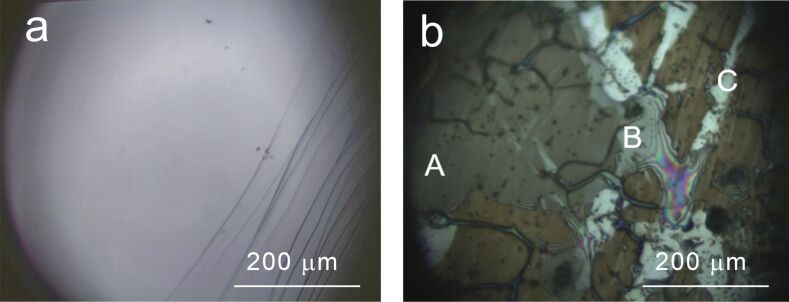
Figure 2.
Optical microscopy image (magnification 50×) acquired ex situ on a) pristine graphite and b) graphite after 15 CVs from 0.3 to 1.6 V. Three different areas are observed: a) a thick brown film is recognized, b) a thinner region of the film where interference fringes are clearly visible, C) areas where no modifications can be observed.

