Figure 5.

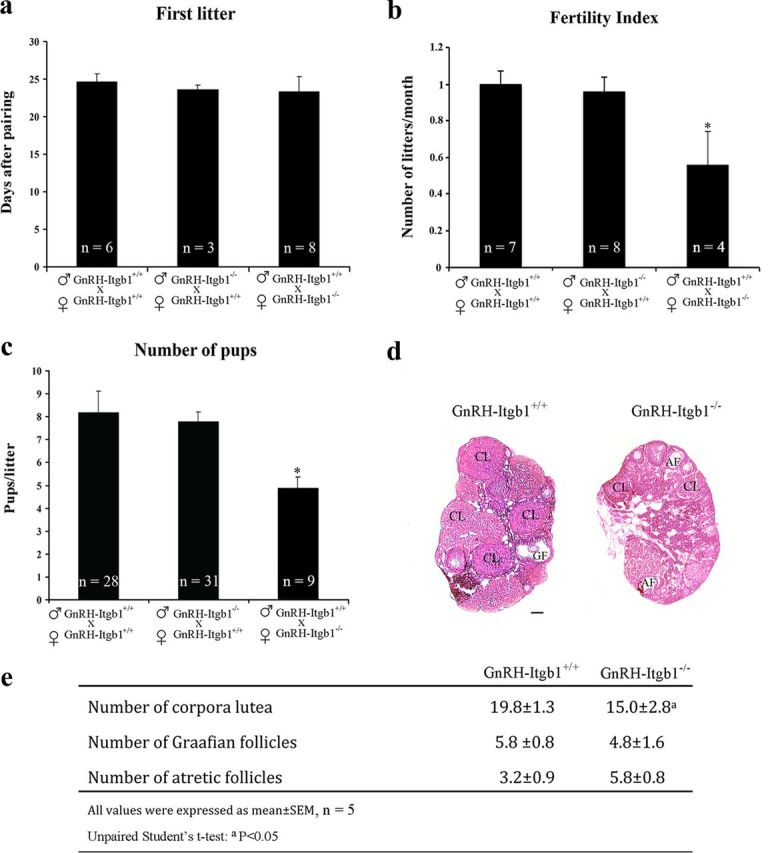
Female GnRH-Itgb1−/− mice exhibit impaired fertility. Fertility in GnRH-Itgb1+/+ and GnRH-Itgb1−/− female mice. Matings were performed for 90 d. a, The latency to first pregnancy was not affected in any group analyzed. b, The total number of litters per female was significantly reduced in conditional mutant female mice compared with control females mated with either GnRH-Itgb1−/− or GnRH-Itgb1+/+ males. c, Conditional mutant female mice gave birth to a reduced number of pups per litter compared with control females. Data are represented as means ± SEM (n, number of animals; *p < 0.01, Fisher 's least significant difference post hoc analysis). d, Morphological analysis of ovaries from 3- to 5-month-old control (GnRH-Itgb1+/+; n = 5) and mutant mice (GnRH-Itgb1−/−; n = 5). Ovary sections (5 μm thick) were stained with hematoxylin-eosin. In GnRH-Itgb1−/− females, the ovaries displayed a greater number of atretic follicles (AF) and a relative paucity of corpora lutea (CL), when compared with the ovaries of control littermates in which follicular development was normal. GF, Graafian follicles. e, The numbers of corpora lutea, Graafian follicles, and atretic follicles were quantified in the ovaries of CNTR and KO mice. Data are represented as means ± SEM. ap < 0.05, unpaired Student's t test. Scale bar, 100 μm.
