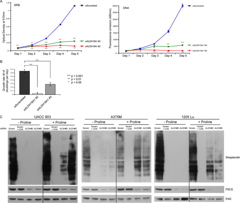
Fig. 5. siRNA-mediated knockdown of P5CS protein levels decreased cell growth rate and protein synthesis.

(A) UACC 903 cells were transfected with siRNA targeting ALDH18A1 or a scrambled control. Cell viability was measured by an SRB assay or Hoechst stain DNA assay for consecutive days (N=6). Points; average, ±SEM. *p<0.05, **p<0.01, ***p<0.001 (B) Optical density (O.D.) values from the SRB and DNA assays representing the exponential growth phase of cells were input into a doubling time calculator from http://www.doubling-time.com and the calculated growth rate values were plotted (N=5). Bars; average, ±SEM. (C) UACC 903, A375M, and 1205 Lu cells transfected with siRNA targeting ALDH18A1 or a scrambled control were subjected to methionine starvation followed by azidohomoalanine (AHA) incubation at 3 (UACC 903), 2 (A375M), or 8 (1205 Lu) days post transfection. Cycloheximide (CHX) served as a positive assay control. Biotin-alkyne was attached to newly synthesized proteins containing AHA before performing western blotting. Membranes were probed with streptavidin-HRP to identify the extent of new protein synthesis and P5CS primary antibody used to confirm protein knockdown. ERK2 primary antibody serves as a control for equal protein loading.
