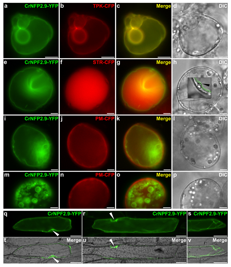
Figure 4. Overexpression of CrNPF2.9-YFP in C. roseus and onion cells.
C. roseus cells were transiently co-transformed with the plasmid expressing CrNPF2.9-YFP and the plasmid encoding the tonoplastic AtTPK1 (TPK)-CFP marker (A-D), the plasmid encoding the vacuolar localized strictosidine synthase (STR-CFP) marker (E-H) or the plasmid containing the plasma membrane targeted aquaporin PIP2A (PM-CFP) marker (I-P). Co-localization of the fluorescence signals appears in yellow when merging the two individual (green/red) false color images (C, G, K, O). Inset in panel H highlights the YFP labelling on the inner side of plastids (white arrowhead) indicating a tonoplastic localization. Cell morphology is observed with differential interference contrast (DIC) (D, H, L, P). Bars, 10 μm Onion cells were transiently transformed with the plasmid expressing CrNPF2.9-YFP (Q-V) and fluorescent signals (Q, R, S) were merged with cell morphology observed with DIC (T, U, V). Black arrowheads highlight the YFP labelling on the inner side of nucleus indicating a tonoplastic localization. Bars, 50 µm.

