. 2016 Dec 23;5:e21198. doi: 10.7554/eLife.21198

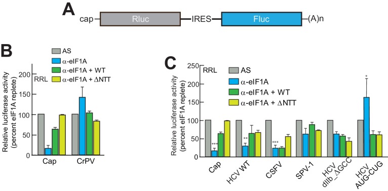
Figure 3. eIF1A depletion inhibits translation from the HCV IRES.

(A) Cartoon showing design of bicistronic dual-luciferase reporter constructs used in the experiments of panels (B–C). (B) Effect of depletion of eIF1A using the aptamer followed by add-back of WT full-length eIF1A (WT) or an N-terminally truncated mutant (ΔNTT) on translation from the reporter shown in panel (A). Experiments were done in RRL. This panel shows the effect on the Rluc (Cap) and a control CrPV IRES. (C) Same as panel (B) but with WT HCV, CSFV, and SPV-1 IRESs, plus two HCV IRES mutants shown in Figure 1B. For panels (B and C), activity in lysate treated with AS control aptamer was set at 100%. Error bars represent averages ±SEM of ≥3 independent experiments. Statistical significance shown by: *p<0.05, **p<0.01, ***p<0.001.

DOI: http://dx.doi.org/10.7554/eLife.21198.006

Figure 3.

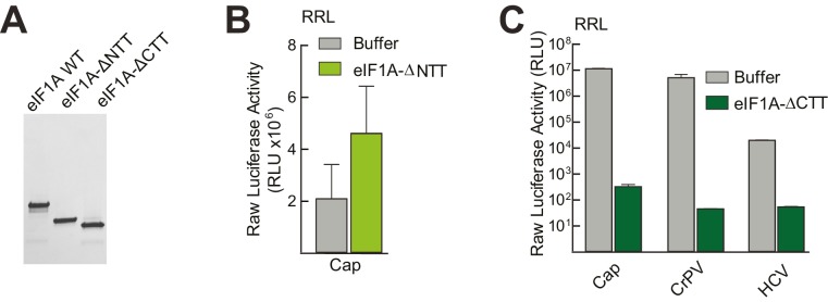
Figure 3—figure supplement 1. Purity of WT and mutant eIF1A and effects in untreated lysate.

Figure 3—figure supplement 1.

(A) Recombinantly expressed and purified WT, N-terminally truncated (ΔNTT), and C-terminally truncated (ΔCTT) human eIF1A shown on a Coomassie-stained SDS-PAGE gel. (B) Translation assays in RRL using a cap-driven message (Figure 3A) with or without the addition of ΔNTT, without aptamer treatment. (C) Translation assays in RRL showing potent translation inhibition by adding ΔCTT. Monocistronic reporters with cap-, CrPV IRES-, or HCV IRES-driven translation are shown. Translation level is shown as raw light units from Fluc. Error bars represent averages ±SEM of ≥3 independent experiments.