
Figure 16. Example of in vivo Ca2+ imaging data collected from superficial, subcortical, and deep brain regions.
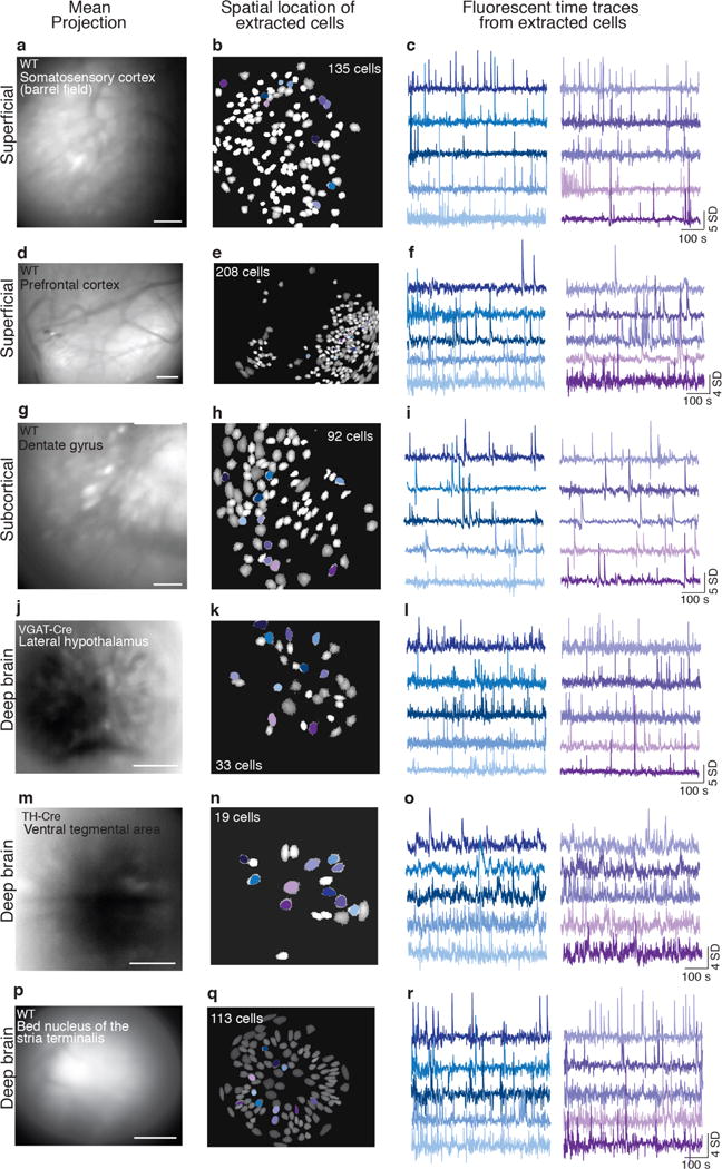
The protocol described here can be adapted for successful in vivo recordings of Ca2+ transients within superficial (a–f), subcortical (g–i), and (j–r) deep brain regions of freely behaving mice. All examples are from one 10-min acute recording session (1 mouse/brain region, data acquired @15 Hz, temporally down sampled to 5 Hz, spatial bin of 4). A 1mm diameter lens was used to image Ca2+ signals in superficial and subcortical areas, while a 0.5 mm diameter lens was in deep brain regions. The left column shows digital images generated by creating a mean intensity projection of fluorescence activity across the entire recording session, the middle column shows the spatial location of optically detected GCaMP expressing cells, and the right column shows activity traces (variation from the mean fluorescence signal over time) of cells outlined in the corresponding spatial map. Spatial maps and activity traces were extracted with PCA/ICA analysis (Mosaic analysis software, Inscopix). (a–c) Somatosensory cortex neurons were targeted by injecting AAVdj-CamkII-GCaMP6f (Titer: 5.6 × 10¹², 1:8 dilution, UNC vector core) into the barrel field region of an adult male wild-type (WT) mouse. (d–f) Prefrontal cortex neurons were targeted by injecting AAV-DJ-CamkIIa-GCaMP6s into the prefrontal cortex of an adult male WT mouse (Titer: 5.3 × 10¹², 1:6 dilution, UNC vector core). (g-i) AAV-DJ-CamkIIa-GCaMP6m was injected into the dentate gyrus of WT male mice (Titer: 7.0 × 10¹², 1:1 dilution, Stanford). (j–l) GCaMP6m was targeted to GABAergic neurons within the lateral hypothalamus by injecting AAVdj-EF1a-DIO-DIO-GCaMP6m (Titer 9.7 × 10¹², 1:2 dilution, Stanford) into adult male VGAT-Cre mice. (m–o) Dopamine neurons within the ventral tegmental area (VTA) of an adult male mouse were targeted by injecting AAVdj-EF1a-DIO-DIO-GCaMP6m (Titer 9.7 × 10¹², 1:2 dilution, Stanford) into a TH-Cre mouse. (p–r) Bed nucleus of the stria terminalis (BNST) neurons were targeted by injecting AAV-DJ-CamkIIa-GCaMP6s (Titer: 5.3 × 10¹², 1:6 dilution, UNC vector core) into wild-type mouse. Notice that deep brain regions differ from cortical regions in image clarity. Injection volumes for all regions were 300 μl. Scale bar = 125 μm. All procedures were approved by UNC IACUC.