
Figure 8. Removal of connective tissue and muscle.
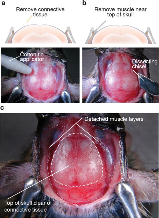
(a) To reduce movement noise during imaging sessions, remove connective tissue by rubbing a cotton tip applicator on the top surface of the skull. Digital image below shows example of skull in which connective tissue has been removed by gently rubbing a cotton tip applicator on the top of the surface. (b) To further reduce the likelihood of movement artifacts, use a dissecting chisel to cut away muscle and tendons making contact with the top edge of the skull. Digital image below shows specific area of the skull where the dissecting chisel should be used to remove muscle. (c) When finished, the surface of the skull should be clear of connective tissue and the muscle retracted from the portion of the skull where dental cement will be applied during the head cap procedure (dotted outline on skull). All procedures were approved by UNC IACUC.