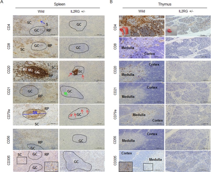
Figure 2. Analysis of T-, B-, and NK-specific biomarker expression patterns in spleen (A) and thymus (B).
T cells (CD4 and CD8), B cells (CD20, CD21, and CD79a), NK cells (CD56 and CD 335) positive signal in WT-derived spleen tissues were strongly detected, whereas a few CD20 and CD79a positive cells in mIL2RG+/Δ69-368 KO pigs, which is a critical factors for B cell development, were detected. However, biomarkers expression of T and NK cells in thymus of mIL2RG+/Δ69-368 KO pigs were not detected. GC, germinal center; RP, red pulp; S, splenic sinusoids; M, mantle; SC, splenic cords; SN, splenic nodule.

