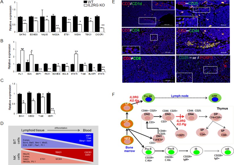
Figure 4. Comparison of B-, T-, and NK-cell specific gene expression by using RT-qPCR and immunofluorescence analysis in WT and mIL2RG+/Δ69-368 KO pig.
mRNA expression of NK A., B., and T C. cells development related genes in mIL2RG+/Δ69-368 KO pig-derived spleen, thymus, and lymph node were significantly down-regulated, whereas expression of PU.1, E2A, and STAT3 encoding early B cell development related genes were up-regulated. D. Cartoon showing major events of T/B/NK cell development. Developing thymocytes, which enter into thymus and migrate into blood, show differential gene expression profiles that modulate the discrete stages of the T/B/NK-cell development. E. To identify T cell developmental stages in mIL2RG+/Δ69-368 KO pigs-derived thymus, CD3 positive cells were co-localized with CD1d or CD4, CD8, CD25, and CD 44 antibodies. Also, CD25 positive cells are colocalized with FOXP3 antibody. F. T cell developmental blocks largely rely on data from targeted mutation studies of mIL2RG+/Δ69-368 KO pigs.

