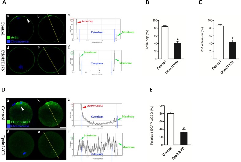
Figure 5. Loss of Episn2 results in the inactivation of Cdc42 in oocytes.
A. mRNAs encoding wild type (control) and dominant-negative mutant of Cdc42 (Cdc42T17N) were injected into fully grown oocytes to evaluate actin cap by Phalloidin (green) labeling. Chromosomes were stained with Hoechst 33342 (blue). B., C. Quantitative analysis of control and Cdc42T17N oocytes with intact actin cap and extruded polar body. D. EGFP-wGBD mRNAs were injected into control and Epsin2-KD oocytes to trace the active Cdc42 (green) during meiosis, and chromosomes were stained with Hoechst 33342 (blue). Representative confocal sections are shown. Arrowhead denotes the position of the polarized EGFP-wGBD. Right graphs show the fluorescence intensity profiling of active Cdc42 in oocytes. Lines were drawn through the oocytes, and pixel intensities were quantified along the lines. E. Quantitative analysis of control (n = 80) and Epsin2-KD oocytes (n = 75) with polarized EGFP-wGBD signals. Data are presented as mean± SD from three independent experiments. *p < 0.05 vs. controls. Scale bar, 20μm.

