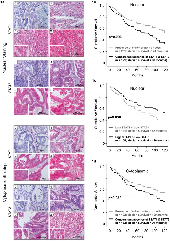
Figure 1. Lower median survival in patients with concomitant absence of nuclear STAT1 and STAT3.

Examples of patient TMAs with nuclear STAT1 and STAT3 staining, showing different levels of STAT expression. a. Immunohistochemical staining was scored by two board certified pathologists as negative (score 0), weak (score 1), moderate (score 2) or strong (score 3). Patients show reduced survival upon concomitant absence of b. nuclear and c. cytoplasmic STAT1 and STAT3. d. Higher nuclear STAT1/STAT3 ratio correlates with increased patient survival.
