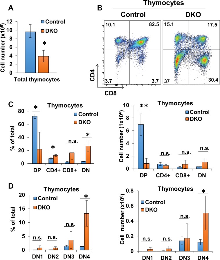
Figure 3. CD4-Cre induced CBL/CBL-B deletion leads to altered thymocyte development.
(A) Mean values of cell numbers from thymuses of 10-week old Control and DKO mice; n = 3. (B) Representative dot plot FACS analysis of anti-CD4 and anti-CD8 stained thymocytes. (C) FACS analysis of CD4CD8 DP, single-positive, and DN thymocyte populations for percentage of total thymocytes (left) and cell number (right); n = 3. (D) Flow analysis of DN thymocyte populations for percentage of total thymocytes (left) and cell number (right); n = 3. DN cells are gated (DN1: CD117+ CD44+ CD25−, DN2: CD117+ CD44+ CD25+, DN3: CD117− CD44− CD25+, DN4: CD117− CD44− CD25−). Data shown are mean +/− SD. ns, p ≥ 0.05; *p ≤ 0.05; **p ≤ 0.01; ***p ≤ 0.001; ****p ≤ 0.0001.

