
Figure 5. Bevacizumab reduces Hs294T cell motility and in vivo metastases formation.
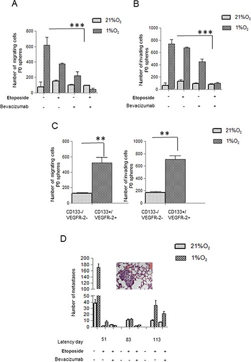
(A, B) P0 spheres from Hs294T, treated with or without Etoposide/Bevacizumab (50 μM) and/or Bevacizumab (250 ng/ml), were disaggregated and cells were analyzed for migration (left) and invasion (right) assay by Boyden Chamber. (C) CD133+/VEGF-R2+ and CD133−/VEGF-R2- sub population were analyzed for cell migration and invasion. (D) Hs294T cells were treated in hypoxic condition with Etoposide (50 μM) alone or with Etoposide (50 μM) plus Bevacizumab (250 ng/ml) for 24 h. 1 × 10⁶ melanoma cells treated as above were injected into the lateral tail vein of SCID bg/bg mice (n = 5 per group) and number of lung macro and micrometastasis was evaluated at different times from injection (e.g. latency). Lungs were inspected for macrometastases using dissecting microscope, and then fixed overnight at 4C in formalin (5% in PBS) for histological analyses of micrometastases photo of micrometastasis of mice injected with hypoxic Hs294T cells treated with Etoposide. Untreated mice were used as control and they all died at the 51st day of observation **p < 0.001. **p < 0.001, ***p < 0.0001, n = 3.