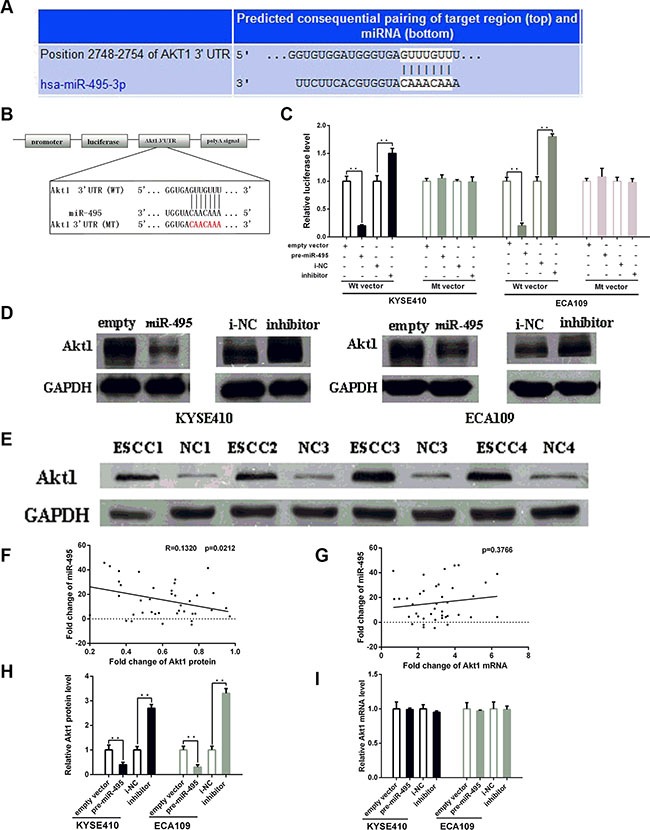
Figure 3. MiR-495 inhibits Akt1 expression by binding its 3′-UTR.

(A) Representations of the predicted target site for miR-495 in the Akt1 mRNA 3′ -UTR. (B) Sequence of the miR-495 binding sites within the human Akt1 3′-UTR and a schematic diagram of the reporter constructs showing the Akt1 3′-UTR sequence (WT) and the mutated sequence (MT). (C) Direct binding of the miR-495 to Akt1 3′-UTR. KYSE410 and ECA109 cells were transfected using a firefly luciferase reporter containing either wild-type (WT) or mutant (Mut) miR-495 binding sites in the Akt1 3′-UTR with miR-495 overexpression or knockdown. 24 after transfection, the cells were assessed using a luciferase assay kit. The results are displayed as the ratio of firefly luciferase activity in the miR-495-transfected cells to that in the control cells. (D) Representative Western blot of Akt1 protein levels in KYSE410 and ECA109 cells after transfection. (E) Representative Western blot of paired tumor (ESCC) and normal adjacent tissues(NC). (F) Pearson's correlation scatter plot of the fold change of miR-495 and Akt1 protein. (G) Pearson's correlation scatter plot of the fold change of miR-495 and Akt1 mRNA. (H) Quantitative analysis of the Akt1 protein and levels in KYSE410 and ECA109 cells after transfection. (I) Quantitative analysis of the Akt1 mRNA and levels in KYSE410 and ECA109 cells after transfection.
