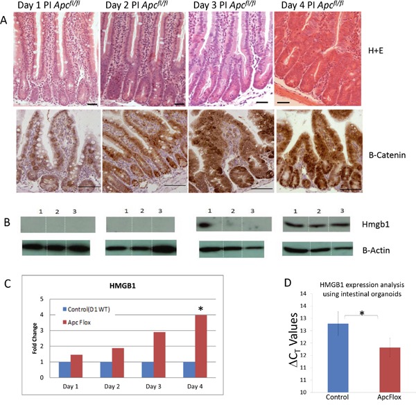
Figure 1. Accumulation of intestinal Hmgb1 following the loss of Apc.

A. H+E stained intestine and β-catenin IHC following the conditional loss of Apc. Days post induction are indicated at the top. Elevated nuclear accumulation of β-catenin occurs from day 3 PI onward. B. Western Blot analysis of HMGB1 levels on triplicate epithelia cell extracts from each time point. Elevation of HMGB1 was first seen at day 3 PI. C. qRT-PCR of Hmbg1 expression levels presented as relative fold change within epithelia cell extracts compared to day 1 WT demonstrates a significant (*) induction 4 days PI (determined using Whitney U test of ΔCT values at P < 0.05). D. qRT-PCR of Hmbg1 expression levels presented as ΔCT values from control (wildtype) and Apc deficient intestinal organoid cultures. The values shown represent a 1.9 fold over-expression of Hmgb1 in the Apc deficient organoids.
