
Figure 6. The IRE1α inhibitor induced cisplatin-mediated apoptosis in cancer stem-like cells.
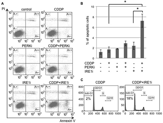
Sphere-forming cells were untreated (control) or treated with 20 μM cisplatin (CDDP) and/or 1.5 μM GSK 2606414 (a PERK inhibitor: PERKi) or 7 μM 4μ8C (an IRE1α inhibitor: IRE1i) for 72 hours. A. After the treatment, cells were subjected to PI/Annexin-V staining and analyzed by flow cytometry. B. A quantitative analysis of PI-negative/Annexin-V-positive apoptotic cells showed that the proportion of apoptotic cells in sphere-forming cells was clearly increased by the IRE1α inhibitor combined with cisplatin treatment. The values shown represent the means ± SEM (*p < 0.05). C. After being treated, cells were fixed, stained with propidium iodide, and the sub-G1 population was calculated by flow cytometry. The cisplatin+IRE1α inhibitor treatment induced apoptosis in sphere-forming cells.