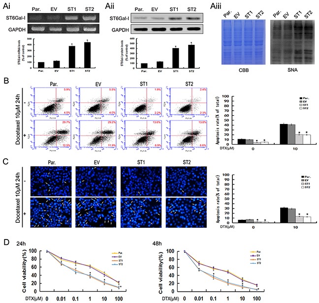
Figure 4. Upregulation of ST6Gal-I increases the survival rate of Huh7 cells and protects Huh7 cells from docetaxel-induced apoptosis.

Ai-Aiii. ST6Gal-I was stably over-expressed in Huh7 cells, the mRNA (Ai) and protein (Aii) levels of ST6Gal-I were determined by RT-PCR and Western-blot assays and normalized for GAPDH (*P<0.05). The α2,6-linked sialic acid (Aiii) levels were determined by SNA lectin staining. Coomassie Brilliant Blue (CBB) staining was used to normalize the protein amounts. B. The rates of apoptosis were determined by flow cytometry analysis of Annexin V-FITC/PI. Results are representative of three independent experiments (*P<0.05). C. Representative images of DAPI staining. Results are representative of 10 different fields (×100) from three independent experiments (*P<0.05). D. Cell viability following docetaxel treatment was detected by CCK8 assay. Note that the (1, 10, 100μM for 24h and 0.1, 1, 10, 100μM for 48h) measurements were significantly different (*P<0.05). Results represent the mean +/− SD of triplicate wells and are representative of at least three independent experiments. Par., Huh7; EV, Empty vector transfectants; ST1 and ST2, pcDNA3.1/ST6Gal-I vector transfected stable clones.
