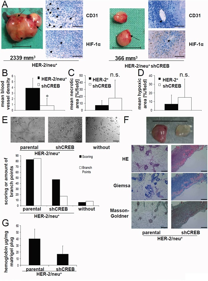
Figure 1. Link of decreased tumorgenicity of CREB-deficient HER-2/neu+ cells and reduced angiogenesis, but enhanced hypoxic areas.

A. DBA-1 mice were injected with parental or CREB-deficient HER-2/neu+ cells as described in Materials and Methods and tumors were removed after 42 days. Representative photos of parental and CREB-deficient HER-2/neu+ tumors are shown. The arrows indicate the blood vessels on the tumor surface. The tumor volume is given. The bar represents 1 cm (left). 5 μm slices of paraffin-embedded tumors were stained with the indicated primary antibody followed by an anti-rabbit secondary antibody. The detection was performed with the peroxidase substrate DAB. Slides were counterstained with methylene blue. The arrow heads indicate the blood vessels. The bar represents 100 μm; Magnification: 40x (right). B. The blood vessel density of the tumors was analysed by counting vessel structures in the anti-CD31 mAb-stained samples (see 1A). Bars represent mean values from four samples/group with four counted fields/sample. C. The necrotic area was analysed in the HE-stained samples. Bars represent mean values from four samples/group with four counted fields/sample. D. The hypoxic area was analysed in the anti-HIF-1α-stained samples. Bars represent mean values from four samples/group with four counted fields/sample. E. 1×10⁴ HUVEC/well were seeded in a 96 well plate on polymerized growth factor reduced matrigel. 100 μl/well fresh medium or cell conditioned medium was added and the cells were incubated for 16 h by 37°C. The morphology of the HUVEC under these distinct culture conditions was compared (left) and the mesh-like structures were quantified (right) as described by Zhang [51]. The bar represents 80 μm; Magnification: 10x. F. 1×10⁵ parental and CREB-deficient HER-2/neu+ cells resuspended in matrigel were injected into the flank of female DBA-1 mice (n = 8). 7 days after injection the mice were killed and the removed matrigel plugs were photographed. The bar represents 1 cm (up). 5 μm slices of the matrigel plugs from parental and CREB-deficient HER-2/neu+ cells were stained as indicated. The bar represents 100 μm; Magnification: 10x (down). G. Matrigel plugs from parental and CREB-deficient HER-2/neu+ cells were homogenized and their hemoglobin content was analysed as described as in Material and Methods. The bars represent the hemoglobin concentration of each plug from mice injected with the indicated cell line normalized to the weight of the plug. Data demonstrate the results of one out of two independent experiments (with five Matrigel plugs in each experiments) regarding the hemoglobin content/plug from parental and CREB-deficient HER-2/neu+ cells.
