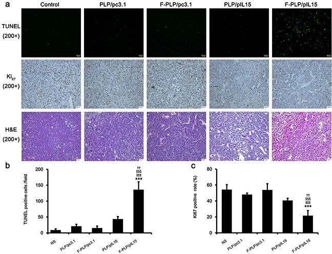
Figure 4. Antitumor mechanisms of F-PLP/pIL15.

a. Representative tumor tissue sections following the TUNEL assay, Ki67 staining and hematoxylin-eosin (H&E; scale bars = 50 μm). b. and c. Tumor cell apoptosis and proliferation were assessed by counting the number of TUNEL-positive cells per field and the Ki67-positive index rate (three high power fields per slide). F-PLP/pIL15 was superior to the controls in increasing tumor apoptosis and inhibiting tumor cell proliferation (*P < 0.05, **P < 0.01, versus NS;‡P < 0.05, ‡‡P < 0.01, versus PLP/pc3.1; §P < 0.05, §§P < 0.01, versus F-PLP/pc3.1; †P < 0.05, versus PLP/pIL15; mean±SD, n = 3).
