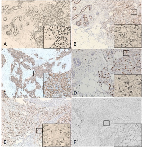
Figure 1. Immunohistochemistry(IHC) Images of ER, PR, Her-2, Ki67 and P53 (×100; ×400).

A. Estrogen receptor (ER) positive; B. Progesterone receptor (PR) positive; C. Human epidermal growth factor receptor 2 (HER2) positive; D., E. Immunoreactions of Ki-67 and p53 were presented through nuclear staining; F. Negative Immunoreactions.
