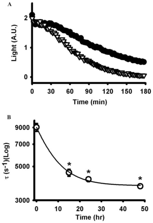
Fig. 4.
Incubation with ATPγS increased ecto-ATPase activity. A, cells exposed to 100 μM ATPγS for 48 h (gray triangles) hydrolyzed 1 μM ATP more rapidly than untreated control cells (black circles). Intermediate preincubations of 15 and 24 h led to intermediate increases in hydrolysis but are not shown here for reasons of clarity. Results show the mean of five to eight wells from a trial representative of three independent experiments. Light levels represent the photons given off with the luciferin/luciferase reaction, an index of the level of ATP present in the bath [in arbitrary units (A.U.)]. B, time constant of decay decreased exponentially with preincubation time. *, p < 0.05 versus no preincubation, n = 5 to 8. In A, error bars are smaller than symbols.

