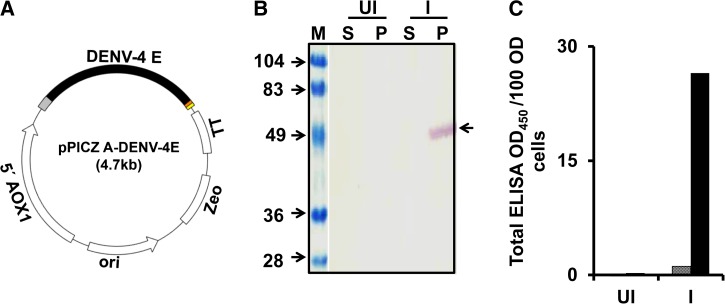
Figure 1.
Expression of DENV-4 E in Pichia pastoris. (A) Schematic representation of DENV-4 E gene cloned in pPICZ-A expression vector with AOX1 promoter (5′ AOX1) upstream and transcription terminator sequence (TT) downstream of it. An Escherichia coli origin of replication (ori) and zeocin resistance (Zeo) marker is also present for efficient replication and transformed clone selection, respectively. (B and C) Evaluation of expression and localization of DENV-4 E in induced P. pastoris. Aliquots of un-induced (UI) and induced (I) cell cultures were subjected to lysis and separated into soluble (S) and membrane-enriched pellet (P) fractions by centrifugation. S and P fractions were evaluated through (B) Western blot and (C) ELISA on Ni-NTA His Sorb plate using DENV-4 E-specific monoclonal antibody (mAb), DENV-4 E88. The blot in panel B is a composite figure, where lane “M” denotes pre-stained markers, the sizes (in kDa) of which are indicated on the left. An arrow on the right of panel B indicates the position of DENV-4 E in the blot. In panel C, the S and P fractions are represented by the grey and black bars, respectively. DENV-4 E = dengue virus serotype 4 ectodomain; ELISA = enzyme-linked immunosorbent assay.

