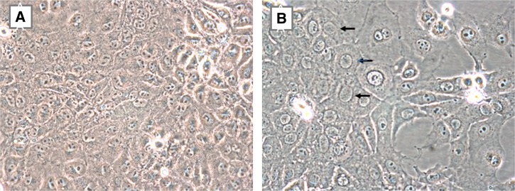
Figure 1.
LLC-MK2 cells. Noninoculated cells (A) appear crowded 13 days postseed; no spaces are present between the cells and the nuclei have prominent nucleoli. (B) Cells 13 days postinoculation with plasma sample four appear different: the nuclei of the infected cells appear larger, lack prominent nucleoli, and have visibly darker nuclear borders (black arrows), and clearings due to detachment of dead cells are evident, as are a few refractile floating dead cells. The virus grew at both 33°C and 37°C; this figure shows virus in cells grown at 33°C. The appearance of the nuclei of the infected LLC-MK2 cells is consistent with HCoV-NL63 infections. Localization to the nucleolus is a common feature of coronavirus nucleoproteins, resulting in the accumulation of cells in the M-phase of the cell cycle (nucleoli are absent in dividing cells), and the formation in syncytia in a fraction of the cells. Moreover, immature HCoV-NL63 particles form in the rough endoplasmic reticulum (RER) surrounding the nucleus, and the dark border surrounding the enlarged nuclei in Figure 1B are likely due to cellular changes in the RER of the infected cells. Representative photomicrographs of cytopathic effects of other viral species are shown in Supplemental Figures 1–3.

