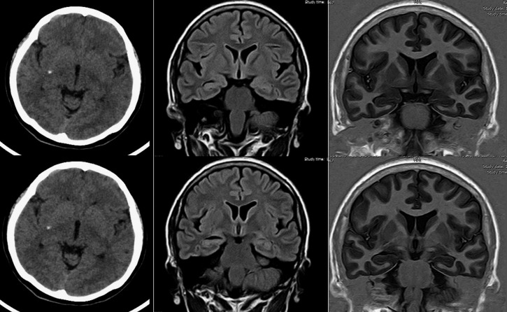
Figure 3.
Neuroimaging of a study participant with unilateral hippocampal atrophy and calcified neurocysticercosis. Plain computed tomography (left column) shows single calcified cysticercus deep in the right side of the brain. Fluid attenuated inversion recovery (center) and T1-weighted inversion recovery magnetic resonance imaging sequence (right) show ipsilateral hippocampal atrophy, characterized by increased width of the choroid fissure and decreased height of hippocampal formation.

