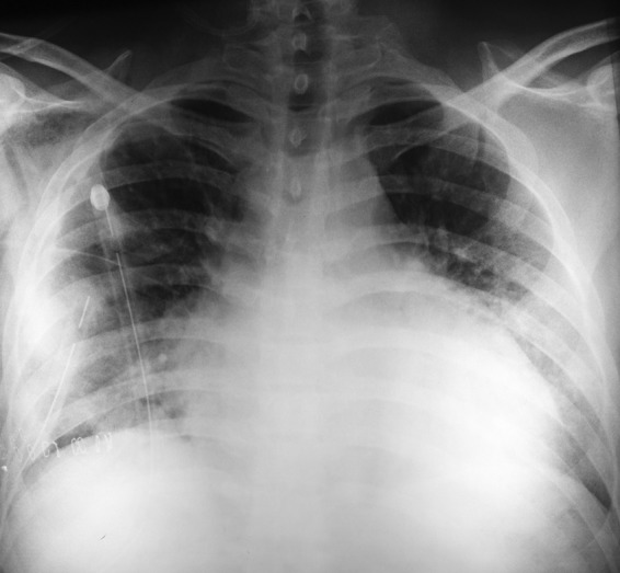
Figure 1.

Anteroposterior chest X-ray performed in year 2011, before the patient's surgery. The image shows bilateral pleural effusions and small nodules in both lung bases. A thoracic tube was inserted in left lung to relieve the patient's symptoms.
