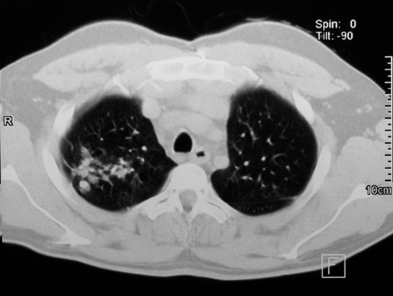
Figure 2.
Axial section thorax computerized tomography scan performed during the patient's hospitalization. Nodular images with thin and regular walls with soft tissue density can be seen in both sides surrounded by areas of scattered frosted glass. Moreover, bronchial dilatation can be appreciated next to the nodular lesions, especially in the right lung.

