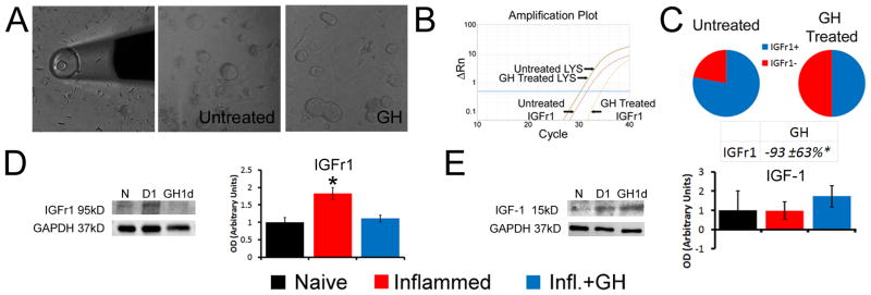
Figure 5. Growth hormone (GH) regulates the expression of insulin like growth factor 1 receptor in vitro and after inflammation in vivo at P14.
Examples of the single cell collection method used for analysis and examples of primary dorsal root ganglion (DRG) cultures treated with or without GH (A). Single cell PCR results from the various culture conditions show that treatment of primary P14 DRG neurons (n=20) with GH significantly reduces the expression of IGFr1 in single cells (B, C). Example of an amplification plot obtained from a cell treated with GH compared to an untreated DRG neuron shows a rightward shift in the Ct value for IGFr1 in the GH treated neuron while the LYS normalization control gene remained constant in each cell (B). In addition to significantly reduced relative expression, the number of cells that express IGFr1 (IGFR1+) at detectable levels in GH treated cultures was also lower than the number of cells containing IGFr1 in untreated DRG neuron cultures (C). One day (D1) after carrageenan induced inflammation of the hairy hindpaw skin, a significant increase in IGFr1 protein is detected in the DRGs; however this is completely prevented in mice treated with GH at P14 (D; n=3–4 for each age). No changes in the ligand IGF-1 however, were detected in the skin among any of the experimental groups tested (E; n=3–4). Examples of IGFr1 and IGF-1 western blots along with their GAPDH are provided in panels D and E. * p < 0.05 vs. naïve (N); One-way ANOVA/Tukey’s post hoc test. Value in C is presented as a percent change from naïve.

