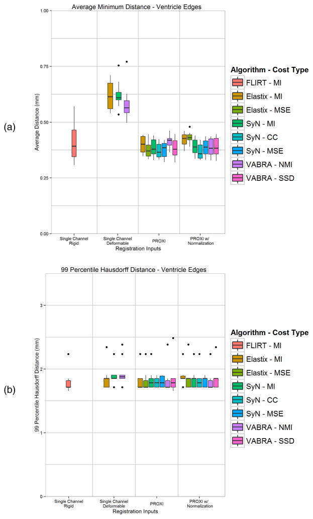
Figure 4.
Boundary comparisons between ventricle edge maps of T1w registration results and the T2w target image using (a) average minimum distance between the edges from both directions, and (b) 99 percentile Hausdorff distance between the edges. Shown are the rigid, single channel MI, and PROXI results using three deformable registration algorithms and their different similarity measures.

