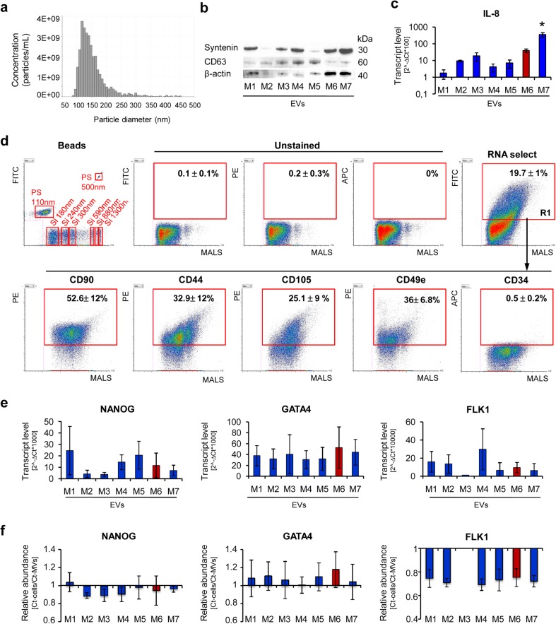
Fig. 4.
Characteristic of EVs derived from UC-MSCs cultured in xeno-free media (UC-MSC-EVs). a Size analysis of EVs using qNano system (Izon Science Ltd). Representative image is shown. b Western blot analysis of selected proteins in UC-MSC-EVs. Three hundred micrograms of protein extracts was used to detect expression of transmembrane (CD63) and cytosolic (syntenin) proteins. Expression of β-actin was used as control. c Transcript level for extracellular protein (IL-8) measured by RT-qPCR in UC-MSC-EVs. d Surface antigen profile of UC-MSC-EVs by high-sensitivity flow cytometry. The EV samples were stained with the SYTO® RNASelect™ Green Fluorescent Cell Stain (Molecular Probes) and selected antibodies labeled with a fluorochrome and further analyzed on an A50-Micro Flow Cytometer (Apogee Flow Systems). The percentage of particles positive for indicated surface marker was analyzed from SYTO® RNASelect™-positive objects (in gate R1). Representative dot plots for M1-EVs are shown. e Analysis of transcript levels for genes involved in the maintenance of pluripotency (NANOG) or differentiation toward cardiac (GATA4) and endothelial lineage (FLK1) performed with the real time PCR method in UC-MSC-EVs. f Relative transcript levels in EVs compared to parental UC-MSCs. Results are shown as mean ± SD. Results were compared with one-way ANOVA and Dunnet’s post hoc test, relative to control conditions (M6). *p < 0.05. UC-MSC umbilical cord-derived mesenchymal stem cells, UC-MSC-EVs extracellular vesicles derived from UC-MSCs, M1 to M7 correspond to the tested media. MALS medium angle light scatter, PS polystyrene calibration beads, Si silicone calibration beads, FITC fluorescein isothiocyanate, PE phycoerythrin, APC allophycocyanin

