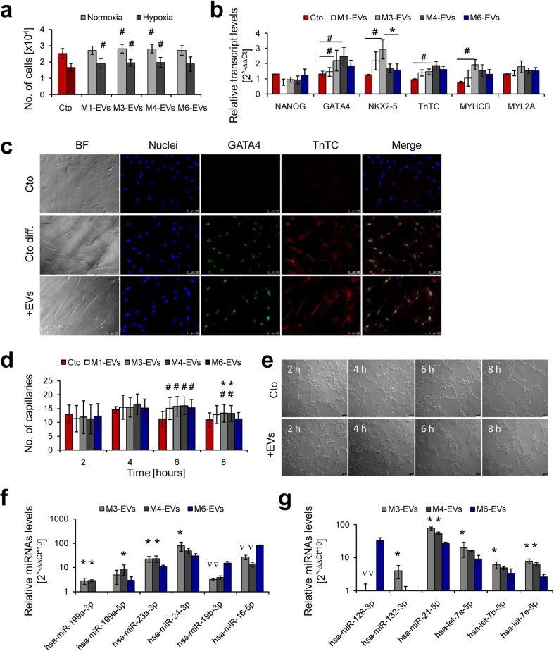
Fig. 5.
Cardiomyogenic and angiogenic potential of xeno-free UC-MCS-EVs in vitro. a Proliferation of cMSCs treated with UC-MSC-EVs (1 μg/103 cells) and cultured 4 days in normal (21 % O2) or hypoxic (1 % O2) conditions, measured with the Cell Counting Kit-8 (Sigma-Aldrich). b Transcript level analysis in cMSCs treated with UC-MSC-EVs (50 μg/5 × 104 cells) and subjected to cardiac differentiation for 7 days. Expression of mRNA was measured using the real time PCR and comparative ∆∆Ct analysis with β-2 microglobulin as endogenous control. c Immunocytochemical staining of cMSCs upon cardiac differentiation for 7 days. Upper panel: undifferentiated control; middle panel: differentiated control (cMSC without treatment with UC-MSC-EVs); bottom panel: cells differentiated after treatment with UC-MSC-EVs. d Capillary formation by human endothelial cells (HUVECs) treated with UC-MSC-EVs (50 μg/5 × 104 cells). Number of capillaries was counted every 2 h during 8-h experiments in five randomly selected microscopic fields. e Microscopic pictures of capillaries formed by HUVECs untreated (upper panel) or treated with UC-MSC-EVs (lower panel) at indicated time points. f Detection of pro-cardiomyogenic miRNAs in UC-MSC-EVs. g Detection of pro-angiogenic miRNAs in UC-MSC-EVs. Representative images are shown for microscopic data. Scale bars indicate 100 μm. For graphical charts: results are shown as mean ± SD. Significant differences in values obtained for cells treated with UC-MSC-EVs and untreated differentiated control (#p < 0.05), as well as between xeno-free and control media (M6 shown as a red column, *p < 0.05) were evaluated by ANOVA. For miRNA analysis: samples statistically enriched or underexpressed (p < 0.05) compared to control M6-EVs are indicated with an asterisk or an inverted triangle, respectively. UC-MSC-EVs extracellular vesicles derived from umbilical cord mesenchymal stem cells, cMSCs cardiac mesenchymal stromal cells, HUVECs human umbilical vein endothelial cells, Cto control, Cto diff. differentiated control, BF bright field (color figure online)

