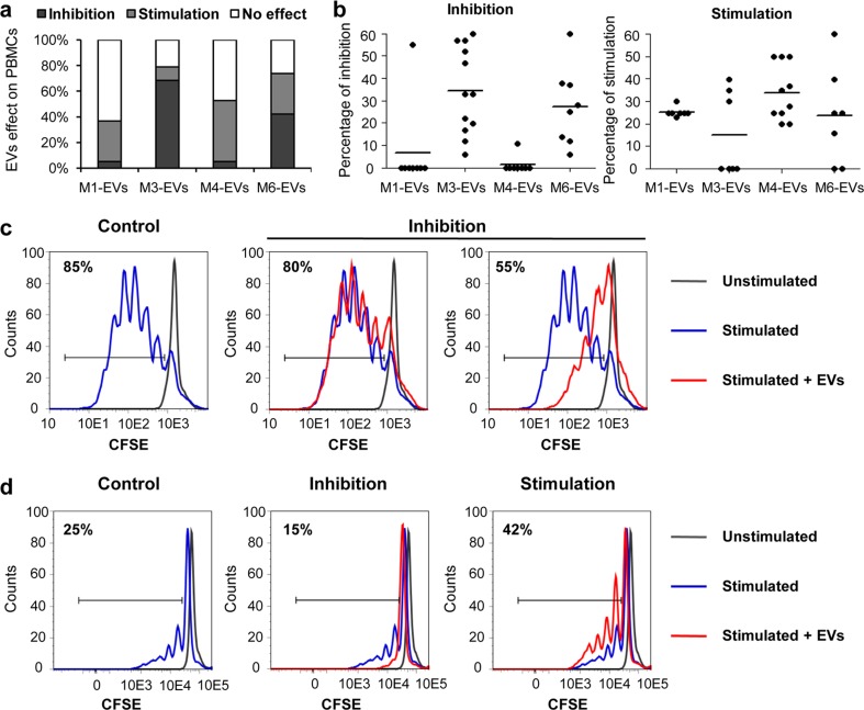
Fig. 6.
UC-MSC-EVs impact on proliferation of peripheral blood mononuclear cells (PBMCs). PBMCs were stained with 25 μM CFSE (Molecular Probes) and stimulated with 50 ng/mL PMA and 1 ng/mL ionomycin (both from Sigma-Aldrich) for 24 h. Next, cells were treated with 100 μg of UC-MSC-EVs for 4 days, and decline in CFSE-positive cells was measured by flow cytometry. a Summary of observed effects exerted by UC-MSC-EVs on PBMCs. Percentage of stimulation, inhibition, or no effect measured in five experiments with the use of four UC-MSC-EV types are shown. b Effects of single UC-MSC-EV samples on proliferation of PBMCs. Every measurement is indicated as a black dot, mean values are shown as horizontal lines. c Inhibition of PBMCs proliferation by two UC-MSC-EV specimens in a sample of a “good responder” to PMA/ionomycin stimulation (>80 % of PBMCs proliferation). Representative histograms are shown. d Dual effect of UC-MSC-EVs on PBMCs proliferation, either stimulation or inhibition, in a sample of a “poor responder” to PMA/ionomycin stimulation (<40 % of PBMCs proliferation). UC-MSC-EVs extracellular vesicles derived from umbilical cord mesenchymal stem cells, PBMCs peripheral blood mononuclear cells, CFSE carboxyfluorescein succinimidyl ester, PMA phorbol 12-myristate 13-acetate

