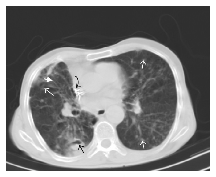
Figure 1.

88-year-old male. Spiral CT scan at the level of aortic root (lung window). Cavitary nodule in right lower lobe (black arrow) along with nodular infiltration in left lower lobe, lingula, right lower lobe, and right middle lobe (white arrows). Note also cylindrical bronchiectasis in right middle lobe (thick white arrow). The hyperdense focus in superior vena cava was related to cardiac pacemaker (curved arrow).
