Figure 3.

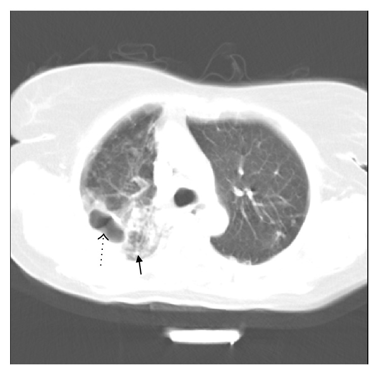
66-year-old female. Spiral CT scan below the level of left pulmonary artery (lung window). There is cavitary consolidation in posterior segment of right upper lobe (dotted arrow) along with adjacent nodular infiltration (thick arrow).

66-year-old female. Spiral CT scan below the level of left pulmonary artery (lung window). There is cavitary consolidation in posterior segment of right upper lobe (dotted arrow) along with adjacent nodular infiltration (thick arrow).