
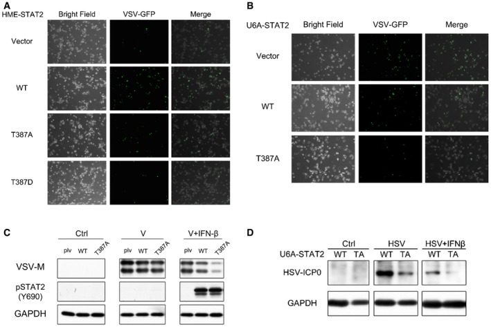
Figure EV2. Phosphorylation of T387 STAT2 inhibits the antiviral effect of IFN‐β in HME and U6A cells.
-
AHME cells expressing wild‐type, T387A, or T387D STAT2 were seeded at 8,000 cells/well. The cells were exposed to VSV for 2 h, with or without pre‐treatment with IFN‐β (100 IU/ml). After 20 h, the expression of GFP from modified VSV was analyzed by microscopy.
-
B, CU6A cells expressing wild‐type or T387A STAT2 were seeded at 8,000 cells/well. The cells were exposed to VSV for 2 h, with or without pre‐treatment with IFN‐β (100 IU/ml). After 20 h, the expression of GFP from modified VSV was analyzed by microscopy (B) and the VSV‐M protein was analyzed by Western blot (C).
-
DU6A cells expressing wild‐type or T387A STAT2 were seeded at 8,000 cells/well. The cells were exposed to HSV for 2 h, with or without pre‐treatment with IFN‐β (100 IU/ml). After 20 h, the HSV ICP0 protein was analyzed by Western blot.