
Figure EV5. ISG induction by IFN‐β in response to dinaciclib treatment and in the absence of STAT1.
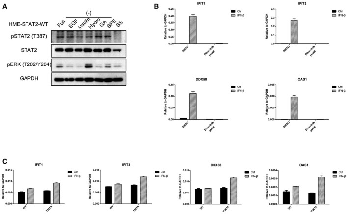
- HME cells expressing wild‐type STAT2 were seeded in complete medium. On the second day, the medium was replaced with fresh medium lacking each component. On the third day, the cells were harvested and total lysates were analyzed by Western blot.
- HME cells were pre‐treated with dinaciclib (4 nM) for 3 h and then treated with IFN‐β (100 IU/ml). Cells were harvested after 4 h, and total RNAs were analyzed by real‐time PCR. Values are the means ± SD from three independent experiments.
- STAT1‐null U3A cells expressing wild‐type (WT) or T387A STAT2 were treated with IFN‐β (100 IU/ml). Cells were harvested after 4 h, and total RNAs were analyzed by real‐time PCR. Values are the means ± SD from three independent experiments.