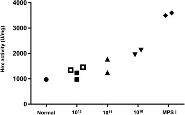
Figure 5.
Normalization of brain hexosaminidase activity in human IDUA tolerant MPS I dogs treated with intrathecal AAV9. Hex activity was measured in samples collected from 6 brain regions (frontal cortex, temporal cortex, occipital cortex, hippocampus, medulla, and cerebellum). The mean activity is shown for 1 normal control dog, 2 untreated MPS I dogs, and the 8 hIDUA tolerant dogs treated with intrathecal AAV9 expressing human IDUA. Open symbols indicate animals tolerized with infusion of recombinant human IDUA. Hex activity was significantly reduced in the high dose cohort compared to untreated controls (p = 0.014, Kruskal-Wallis test followed by Dunn's multiple comparisons test).

