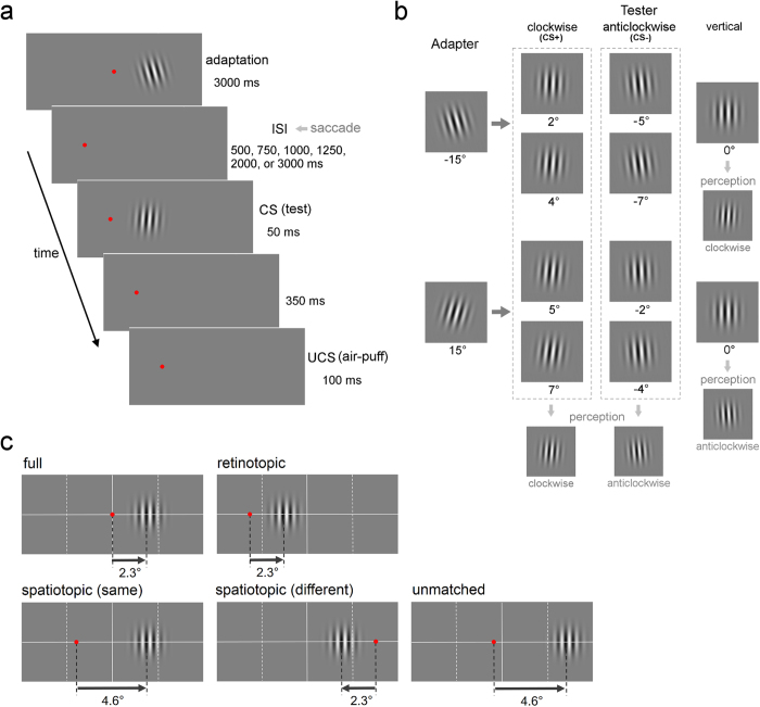
Figure 1. Design of Experiment 1.
(a) Trial sequence in the retinotopic condition. The adapter stimulus was presented for 3000 ms. After the ISI, the test stimulus (CS) was presented for 50 ms. The UCS (air puff) was presented 350 ms after the CS for 100 ms. The FP was moved after the presentation of the adapter stimulus, except for the full condition. The participants were asked to perform a saccade to the new FP as soon as possible. (b) The four types of trials for each CS+ and CS− trial. Adapter stimuli were Gabor patches rotated clockwise and counterclockwise by 15°. Test stimuli (CS) were patches rotated clockwise and counterclockwise by 2°, 4°, 5°, and 7°. For half of the participants, the CS+ was four patches rotated clockwise and the CS− was four patches rotated counterclockwise. The test patches should appear to be tilted clockwise in all four CS+ trials and they should appear to be tilted counterclockwise in all four CS− trials. For the remaining half of the participants, the CS+ and the CS− were reversed. A vertical test patch was also presented after the clockwise and counterclockwise adapters, not followed by the UCS. After differential conditioning is acquired, eyeblink CRs are observed for the vertical patch when it appears to be tilted in the same direction as the CS+ due to TAE. (c) The location of the FP and the test stimulus presented after the adapter stimulus in the five reference frame conditions: the full, retinotopic, two spatiotopic, and unmatched conditions. In one spatiotopic condition, the adapter and the tester were presented in the same hemi-field. In the other spatiotopic condition, they were presented in different hemi-fields.

