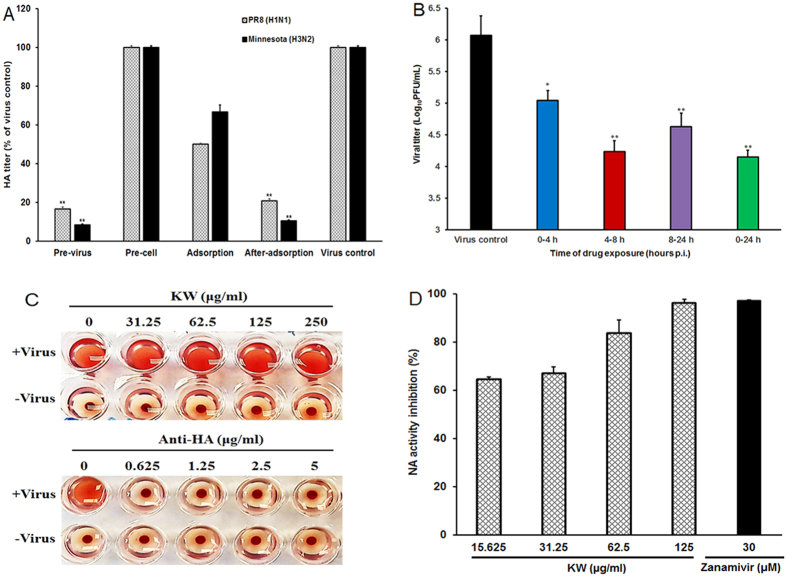
Figure 3. Influence of different treatment conditions of fucoidan on IAV infection.
(A) MDCK cells were infected with Minnesota (H3N2) or PR8 (MOI = 0.1) under four different treatment conditions. (i) Pretreatment of virus: IAV was pretreated with 250 μg/mL of KW at 37 °C for 1 h before infection. (ii) Pretreatment of cells: MDCK cells were pretreated with 250 μg/mL of KW before infection. (iii) Adsorption: cells were infected in media containing 250 μg/mL of KW and, after 1 h adsorption at 37 °C, were overlaid with compound-free media. (iv) After adsorption: after removed unabsorbed virus the infecting media containing 250 μg/mL of KW were added to cells. At 24 h p.i., the antiviral activity was determined by HA assay. Values are means ± S.D. (n = 3). Significance: **p < 0.01 vs. virus control group. (B) PR8 (MOI = 0.1) infected MDCK cells were treated with 250 μg/mL of KW for the specified time period, and then the media were removed and cells were overlaid with compound-free media. Then at 24 h p.i., the cell supernatants were collected and the virus yields were determined by plaque assay. Values are means ± S.D. (n = 3). Significance: *p < 0.05 vs. virus control group. (C) The inhibition effects of KW and anti-HA antibody on IAV-induced aggregation of chicken erythrocytes were evaluated by hemagglutination inhibition (HI) assay. (D) Inactivated PR8 virus was incubated with indicated concentrations of KW or Zanamivir (30 μM), and the NA enzymatic activity was determined by a fluorescent assay. Values are means ± S.D. (n = 4).

