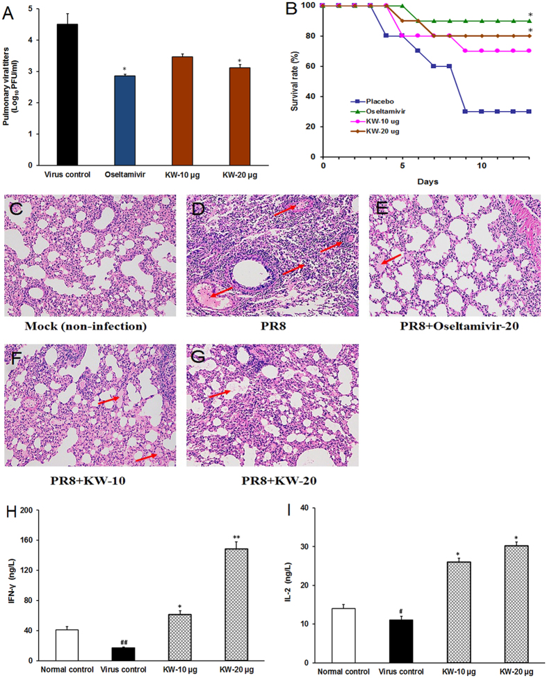
Figure 6. The anti-IAV effects of fucoidan KW in vivo.
(A) Viral titers in lungs. After treatment with KW (10 or 20 μg/day) or placebo (PBS) for 4 days, four mice per group were sacrificed and the pulmonary viral titers were evaluated by plaque assay on MDCK cells. Values are means ± S.D. (n = 3). Significance: *p < 0.05 vs. virus control group. (B) Survival rate. IAV infected mice were received intranasal therapy with KW (10 or 20 μg/day) or placebo once daily for seven days. Results are expressed as percentage of survival, evaluated daily for 14 days. Significance: *p < 0.05 vs. control group (placebo). (C–G) Histopathologic analyses of lung tissues on Day 4 p.i. by HE staining (×10). The representative micrographs from each group were shown. Mock: non-infected lungs; PR8: IAV infected lungs without drugs; PR8 + Oseltamivir-20: IAV infected lungs with Oseltamivir (20 mg/kg/day) treatment; PR8 + KW-10: IAV infected lungs with KW (10 μg/day) treatment; PR8 + KW-20: IAV infected lungs with KW (20 μg/day) treatment. The red arrows indicate the presence of inflammatory cells in the alveolar walls and serocellular exudates in the lumen. (H,I) After treatment of KW (10 or 20 μg/day) for four days, the production of interferon-γ (IFN-γ) (H) and interleukin 2 (IL-2) (I) in spleen tissues was determined by using the ELISA kits for IFN-γ and IL-2. Values are means ± S.D. (n = 4). Significance: #P < 0.05, ##P < 0.01 vs. normal control group; *P < 0.05, **P < 0.01 vs. virus control group.

