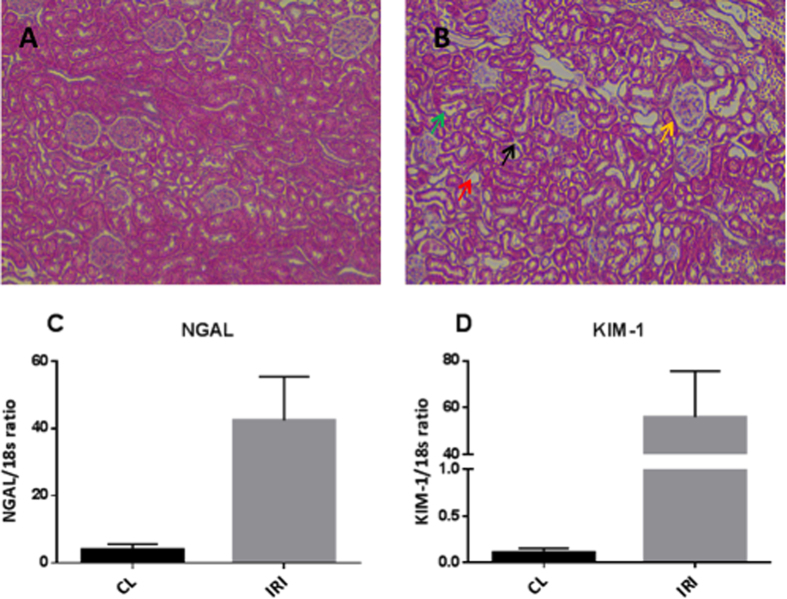
Figure 1. Verification of ischemia-reperfusion injury.
Representative histological sections are shown in (A) a CL kidney showing normal intact tubular cells and glomeruli, and (B) a post-ischemic kidney showing a cellular cast in the tubular lumina (green arrow), complete sloughing of tubular epithelium (red arrow), interstitial edema (black arrow), and glomerular edema (yellow arrow). Magnification 20×, HE stain. The relative expression of injury markers indicated significant upregulation of (A) NGAL (p = 0.0145, n = 6) and (B) KIM-1 (p = 0.0256, n = 6). A paired two-sided Student’s t-test was used to compare the CL and IRI kidneys. Blocks indicate means, while bars indicate the s.e.m. CL = contralateral kidney; HE = hematoxylin and eosin; NGAL = neutrophil gelatinase-associated lipocalin; KIM-1 = kidney injury molecule 1; IRI = ischemia/reperfusion injury.

