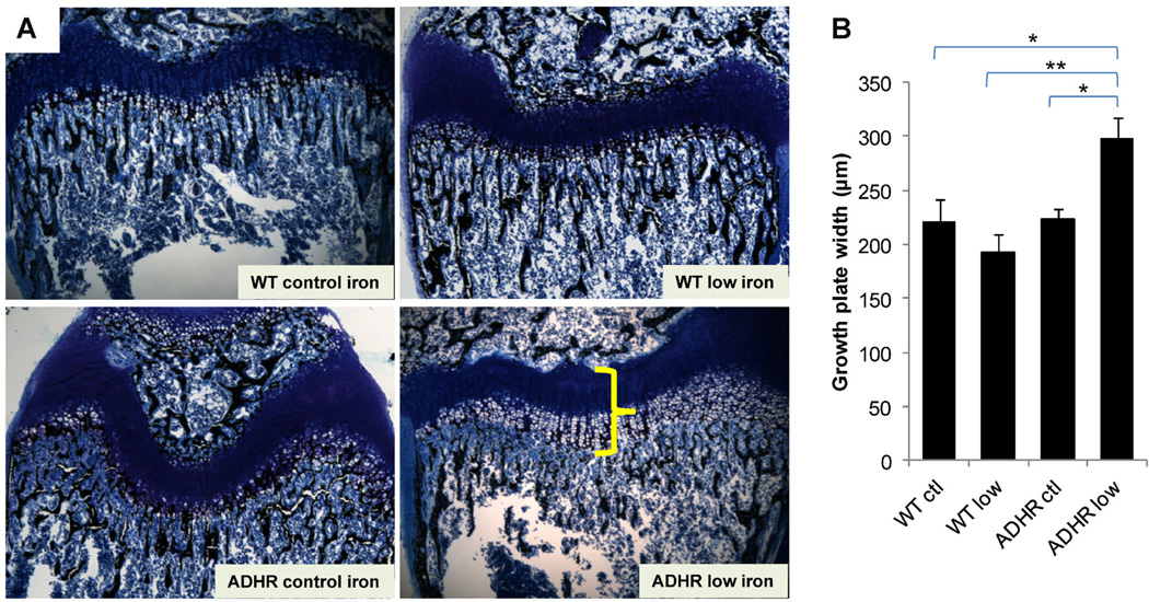
Fig. 4.
Distal femur histomorphometry. (A) Femur distal metaphysis sections were stained with McNeal/Tetrachrome and assessed using histomorphometry. The ADHR low-iron diet mice (lower right panel) had significantly widened growth plates characterized by an expanded zone of calcification (shown by yellow bracket) compared to WT control diet (upper left), WT low-iron diet (upper right panel), and ADHR mice receiving the control iron diet (lower left). (B) Quantification of the growth plate defect demonstrated significant increases in plate width for the ADHR low-iron group (“ADHR low”) versus all other groups (n = 4–5/group: **p < 0.01 versus WT low iron (“WT low”; *p < 0.05 versus WT and ADHR control (“ctl”) iron diet).

